November 26, 2025 by Simonas Bendžius, Center for Physical Sciences and Technology
Collected at: https://phys.org/news/2025-11-copper-nanoparticles-unexpectedly-suitable-ultraviolet.html
Lithuanian researchers at the Center for Physical Sciences and Technology (FTMC), Habil. Dr. Gediminas Niaura and Dr. Martynas Talaikis, together with international colleagues, have for the first time demonstrated that copper is a suitable metal for ultraviolet surface-enhanced Raman spectroscopy (UV SERS)—a highly sensitive method used to study molecular vibrations.
Their findings were published in Advanced Optical Materials and also appear on its back cover. The achievement is significant for many: It opens the door to far more precise detection of low-molecular-weight biochemical compounds and represents a first step toward next-generation early skin cancer diagnostics.
“The field we’ve chosen is still under-explored. Very few researchers in the world work on this,” say the FTMC Department of Organic Chemistry scientists.
An unexpected discovery
First, a brief reminder: Raman spectroscopy is a widely used scientific method in which laser light directed at a material reveals its molecular vibrations, providing researchers with a wealth of information about its properties.
The FTMC physicochemists set an ambitious goal: to adapt one of the most advanced vibrational spectroscopy techniques—surface-enhanced Raman spectroscopy—for practical medical applications, specifically for detecting early-stage skin cancer. Although SERS is commonly used in fundamental and applied biomedical research, it has not been adopted clinically due to insufficient reproducibility and a lack of methodological standardization. Another major limitation is its relatively low selectivity in identifying low-molecular-weight cancer biomarkers.
The question, then, was this: how can the selectivity and reproducibility of SERS be improved? The answer lies in the fact that many biologically important molecules (including cancer markers) absorb ultraviolet (UV) radiation much more strongly than other tissue components. When UV light is used, the Raman signal of these molecules becomes significantly stronger—thereby improving SERS selectivity.
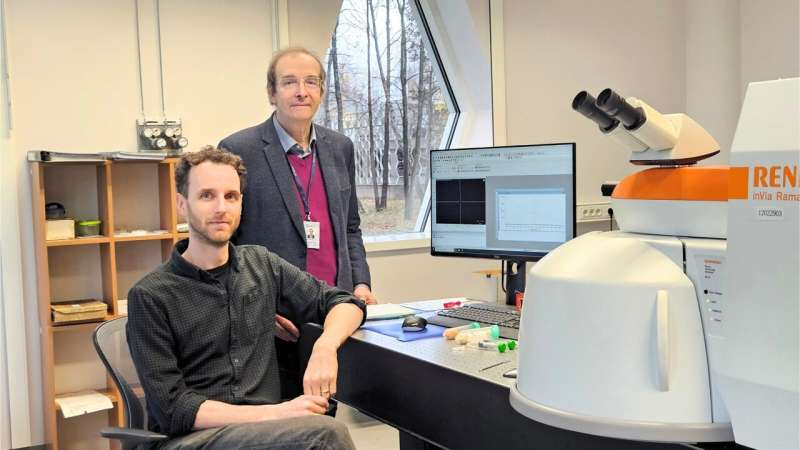
Dr Martynas Talaikis and Habil. Dr Gediminas Niaura. Credit: FTMC
“Our aim is to use UV SERS as a diagnostic method for rapid and reliable detection of cancer-related spectral markers. Ideally, this would be possible even during surgical operations. A new compact device would make this feasible,” explained Gediminas Niaura in January when presenting the group’s project.
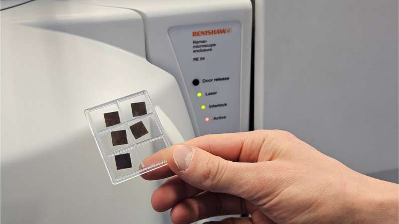
To achieve this, the Lithuanian researchers and their international partners have been developing and testing nanoparticles made of various metals. These particles are essential to SERS: they act as highly sensitive signal amplifiers, enhancing the Raman response of specific biological molecules by millions of times. The idea is simple: nanoparticles are mixed with non-invasively collected skin swab samples and illuminated with UV light. If early-stage cancer markers are present, their spectral fingerprints become clearly visible—allowing diagnosis much earlier than traditional methods.
And now, having launched the project last year, the FTMC researchers have obtained their first promising and unexpected results: Copper nanoparticles turned out to be excellent SERS enhancers under UV laser illumination. This result was surprising because it has long been known that UV light does not induce the electromagnetic field enhancement on copper surfaces that is normally required for the SERS effect. Instead, a different and less-studied mechanism dominates here—chemical enhancement.
The breakthrough came unexpectedly when Dr. Vladimir Sivakov of the Leibniz Institute of Photonic Technology, later a co-author of the paper, arrived at FTMC with nanoparticles produced in his laboratory. “They included bismuth, copper, and other metals. We carried out tests under different irradiation conditions to obtain a reliable SERS signal. Bismuth didn’t work at all, but copper responded remarkably well to UV light,” recalls Dr. Martynas Talaikis.
Molecular motions and ‘fingerprints’
“With copper, we were able to reliably detect low-molecular-weight aromatic compounds such as the nucleobase adenine. Copper particles turned out to be quite stable—we kept them unchanged for a couple of months. As mentioned, the chemical enhancement mechanism on the copper surface proved crucial in this research. When a molecule adsorbs—effectively ‘sticks’—to the metal surface, its electronic structure changes, and this change leads to enhanced interaction with laser light via resonance effects, significantly boosting the signal,” explains Niaura.
“You can shine light on a molecule even without a metal surface: if the laser wavelength matches its absorption, the signal may increase by tens of thousands of times—that’s well-known. But once the molecule binds to a metal, new effects arise, and the results can be even more impressive,” adds the professor.
The FTMC researchers attached adenine—a key biological molecule widely used in laboratories around the world—to the copper surface, allowing their spectral data to be easily compared with international research.
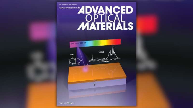
Back cover of the Advanced Optical Materials. Credit: Wiley
How does this look in practice? On the back cover of Advanced Optical Materials we see, at the bottom, a silicon platform covered with copper nanoparticles. The surface appears smooth, but under a microscope it would reveal countless tiny “nanoscale bumps.” At the top, UV laser light is directed at the copper. In the center, the structural formula of adenine and corresponding SERS data are shown.
Interestingly, all molecules have their own unique “fingerprint,” which researchers can read on a computer screen to identify the substance. Each peak in the illustrated SERS spectrum corresponds to a different molecular vibration—demonstrating just how sensitive the method is.
Advanced technology just around the corner?
The ultimate goal of the Lithuanian team is to develop a biosensor capable of rapidly and reliably identifying skin cancer. FTMC is collaborating in this area with Malmö University Professor Dr. Tautgirdas Ruzgas. The envisioned diagnostic procedure would be simple: a moistened sponge would be applied to a suspicious patch of skin, washing off a small number of molecules from its surface. These would then be transferred to a prepared sample containing nanoparticles, illuminated with UV light—and a rapid result would be obtained.
“After publishing our paper, we are now searching for other suitable metals for experiments, but we are also continuing work on copper,” says Prof. Niaura. “We plan to create composite nanoparticles consisting of copper and magnetic components, which would serve a dual function. Magnetism helps control particle deposition, concentration and purification.”
The FTMC scientists emphasize that although much work remains before their idea can be applied clinically, the method is steadily improving—and they have already taken a strong step toward a reliable, stable and cost-effective technology.
More information: Shivani Yadav et al, Copper‐Based Multiwavelength UV Surface Enhanced Raman Spectroscopy, Advanced Optical Materials (2025). DOI: 10.1002/adom.202500078
Leave a Reply