December 5, 2025 by Gerhard Samulat, European XFEL
Collected at: https://phys.org/news/2025-12-ray-spikes-reveal-electron-size.html
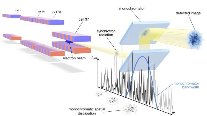
While synchrotron radiation is often thought of as “stable,” the electromagnetic field exhibits pronounced randomly fluctuating distributions both temporally and spatially. These fluctuations encode spatial information about the electron beam that produces the X-rays.
A team led by Andrei Trebushinin and Svitozar Serkez of European XFEL was the first to detect these fluctuations and used them to non-invasively measure the electron beam size along SASE beamlines at each undulator cell. The undulator is the device that forces accelerated electrons to emit X-rays.
The experiment was conducted at the SASE1 beamline of European XFEL using existing equipment: a silicon monochromator and a synchrotron radiation imager. “The short electron bunches from our linear accelerator are the key,” explains Andrei Trebushinin, lead author of the study published in Physical Review Letters. A separate study is published in the journal Physical Review Accelerators and Beams.
The method could in principle also be applied at storage rings, provided sufficient statistics can be collected. But because of the length of the bunches in storage rings, a similar device would require ultra-high-resolution monochromators. “Here, we can just do it with our facility’s monochromator,” Trebushinin says. The information is extracted purely from the statistical structure of intensity fluctuations, similar to the Hanbury Brown-Twiss experiment for measuring the angular diameter of stars that revolutionized stellar astronomy in the 1950s.
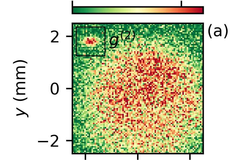
Measurement of intensity patterns at a single undulator cell. The retrieved electron beam size is 16.8 ± 0.4 μm horizontally and 26 ± 0.3 μm vertically. Credit: Physical Review Accelerators and Beams (2025). DOI: 10.1103/31gl-qyk7
The measurements are important for advanced XFEL operations schemes such as attosecond pulse generation, self-seeding, or two-color lasing. The new method enables cell-by-cell diagnosis and eliminates the need for wire scanner installations at various locations. “This is a beautiful experimental demonstration of statistical optics effect applied to synchrotron radiation,” explains Gianluca Geloni, group leader at European XFEL. The research team included collaborators from European XFEL and DESY as part of the free electron laser (FEL) R&D program.
More information: Andrei Trebushinin et al, First Observation of Synchrotron Radiation Spikes for Transverse Electron Beam Size Measurements at a Free-Electron Laser, Physical Review Letters (2025). DOI: 10.1103/z89g-f7j6
Andrei Trebushinin et al, Noninterferometric method for transverse electron beam size diagnostic with synchrotron radiation at a free-electron laser, Physical Review Accelerators and Beams (2025). DOI: 10.1103/31gl-qyk7
Journal information: Physical Review Letters
Leave a Reply