December 5, 2025 by King’s College London
Collected at: https://medicalxpress.com/news/2025-12-ai-brain-scan-tumors-aneurysms.html
A new AI model could help radiologists identify brain abnormalities in MRI scans for all conditions including stroke, multiple sclerosis and brain tumors.
The study, led by researchers at King’s College London and published in Radiology AI, shows how AI could address the growing backlogs due to radiologist shortages as well as an increasing demand for MRIs year on year for over a decade.
These backlogs could result in treatment delays and poorer patient outcomes because MRI scans are vital for diagnosing and monitoring a range of brain conditions such as tumors, strokes and aneurysms.
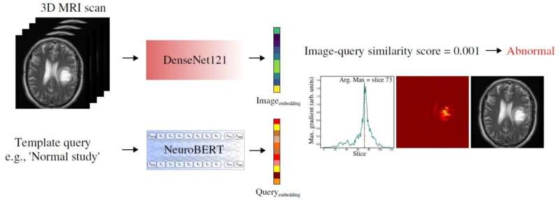
How the AI model was developed
AI could help ease the pressure on radiology departments by triaging scans and increasing reporting speeds.
To do this, the model was first asked to distinguish between ‘normal’ and ‘abnormal’ scans, which it did accurately when compared to assessments made by expert radiologists.
It was then tested on specific conditions—using new MRI scans which weren’t included in the training data—such as a stroke, multiple sclerosis and brain tumors, and was able to recognize these accurately.
Training without manual labeling
Most AI models are currently built with large datasets, manually labeled by expert radiologists—which are expensive and time-consuming to produce.
To overcome this, the team built an AI model that trained itself—without the need for expert radiologists—on over 60,000 existing brain MRI scans using their corresponding radiology reports simultaneously.
“By training the system on scans and the language radiologists use to describe them, we can teach it to understand what abnormalities look like,” explained senior author of the study Dr. Thomas Booth, Reader in Neuroimaging at King’s College London and Consultant Neuroradiologist at King’s College Hospital.
Potential applications and next steps
The researchers also designed the model so that when given a scan or textual query like “glioma,” a type of brain tumor, the system could search and retrieve similar cases, potentially supporting diagnostic review or teaching.
The study indicates that the model could be used at the time of scanning to flag abnormal scans and support clinical decision-making by suggesting findings to radiologists, detecting potential errors in reports, or retrieving similar cases from past examinations. This would speed up diagnoses and reduce reporting delays, helping to improve patient outcomes.
“The next step is to run a randomized multicenter trial across the UK to see how abnormality detection improves workflows in practice. We are pleased to say that this trial will start in hospitals in 2026,” commented Booth.
More information: David A. Wood et al, Self-supervised Text-vision Alignment for Automated Brain MRIAbnormality Detection: A Multicenter Study (ALIGN Study), Radiology: Artificial Intelligence (2025). DOI: 10.1148/ryai.240619
Leave a Reply